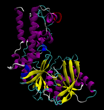


In order to enhance our movie, we are going to change the Material that we are using to add color to the structure. With this coloring method, each secondary structure will be colored by a different color, which highlights each region accordingly. In the ‘Coloring Method’ drop-down menu, select: Secondary Structure Let us change the color to enhance these features. This will change the representation of the protein for a more easier visualization were alpha helices and beta sheets are easier to distinguish. In the ‘Drawing Method’ menu, click and select: NewCartoon Now let us choose a better representation for the protein. This will select only the atoms that are included in the protein. In the ‘Selected Atoms’ field, delete the word ‘all’ write the word protein. To do this, we have to select the protein only. We will use different methods of visual representation for the protein and the ligand. In this window we can adjust the representations of the protein, the ligand and the ions if needed. This will open the ‘Graphical Representations’ window. Close the ‘Molecule File Browser’ and go to: Now that we have loaded the topology and the 5074 frames, we can edit how the system will look. You should see the protein in the main VMD window (the windows with the name ‘VMD 1.9.3 OpenGL Display’). This means that the trajectory file that we are going to load is going to be linked with the topology file, hence the importance to read the prmtop file before the trajectory file.Ĭlick the load button to load the trajectory file. Make sure that the ‘Load files for:’ has the vph.prmtop topology selected. Now click the Browse button and select the trajectory file vph.nc. Load the vph.prmtop topology first! Select the file and click the Load button. With the topology and the trajectory file downloaded, go to:
#ANIMATE SECONDARY STRUCTURE VMD PC#
This tutorial has been prepared in an PC computer running the Linux Ubuntu operating system with VMD version 1.9.3. The topology file is vph.prmtop and the trajectory file is vph.nc. For this tutorial we are going to use a trajectory of a protein receptor that has a bound ligand in the active site. In this tutorial we are going to use the Visual Molecular Dynamics ‘VMD’ visualizer from developed at the University of Illinois at Urbana-Champaign. Movies are always useful to show a simulation.
#ANIMATE SECONDARY STRUCTURE VMD MOVIE#
You want to generate a nice movie from your trajectory data. Making a movie from an AMBER trajectory using VMD


 0 kommentar(er)
